

Blog
15 October 2026
mRNA translation efficiency: How to measure and optimize protein output
For an mRNA therapeutic, getting the RNA into the cell is only part of the challenge. The transcript must also remain stable, engage the translation machinery and produce enough of the intended protein to achieve the desired biological effect.
That makes mRNA translation efficiency a key consideration in RNA therapeutics. Two transcripts can be present at similar levels inside a cell yet produce very different amounts of protein. Differences can arise from the 5′ cap, UTRs, coding sequence, RNA structure, codon usage, nucleotide modifications and the cellular environment. A recent review identifies translation efficiency and mRNA turnover as two critical determinants of therapeutic mRNA productivity [1]. Nature
Measuring RNA abundance alone is therefore not always enough. To understand whether an mRNA is functioning as intended, researchers need to look at what happens at the level of translation.
What is mRNA translation efficiency?
mRNA translation efficiency describes how effectively an mRNA is used by the cellular translation machinery to produce protein. At the transcriptome level, relative translation efficiency is commonly estimated by comparing the Ribo-seq signal for a transcript with its abundance measured by RNA-seq [2].
RNA-seq tells us how much of a transcript is present. Ribosome profiling, or Ribo-seq, adds another layer by sequencing ribosome-protected mRNA fragments and showing where ribosomes are positioned across translated RNAs [2]. PubMed Central (PMC)
For RNA therapeutic development, the practical question becomes not only “Is the mRNA present?” but also “How strongly is it being translated?”
Why translation efficiency matters in RNA therapeutics
Therapeutic mRNA performance depends on several connected steps. The RNA must remain intact, reach the appropriate cellular compartment, recruit the translation machinery and support protein synthesis. Optimizing one step does not guarantee success at the next.
A large-scale study assembled translation-efficiency measurements from 3,819 ribosome profiling datasets covering more than 140 human and mouse cell types [3]. This extensive atlas highlights how translation can vary across biological contexts and provides a framework for studying sequence-dependent translational control. Nature
Translation also changes with cell state. Chothani, Rackham and colleagues combined RNA-seq and ribosome profiling to follow human cardiac fibroblasts as TGFβ1 converted them into myofibroblasts. They found widespread translational effects, identified RNA-binding proteins such as PUM2 and QKI as hubs of post-transcriptional regulatory networks, and showed that these networks were also active in hearts of patients with dilated cardiomyopathy. The authors reported that about one-third of genes were translationally regulated during this transition, a layer of control that RNA-seq alone would have missed.
For therapeutic development, this means translation should ideally be evaluated in a biologically relevant system rather than inferred from RNA abundance or sequence design alone.
What controls mRNA translation efficiency
1. The 5′ UTR and translation initiation
Translation initiation is a major control point in protein synthesis. The 5′ untranslated region can influence ribosome recruitment, scanning and start-site recognition.
Lewis and colleagues quantified ribosome recruitment across more than 30,000 human 5′ UTRs and observed approximately a 200-fold range in translation output [4]. They also identified short regulatory elements that strongly affected translation and showed that N1-methylpseudouridine can alter translation initiation in a sequence-dependent manner [4]. PubMed
Upstream open reading frames (uORFs) add a further layer of control. In a high-resolution map of human translation across six primary cell types and five tissues, Chothani and colleagues detected 7,767 translated small ORFs (smORFs) with translational profiles resembling those of known proteins, many of them cell-type- or tissue-specific.. Notably, changes in the translation efficiency of uORFs and of their downstream main ORFs mostly occurred in the same direction, suggesting that the relationship between uORF and main-ORF translation is more nuanced than simple repression.
A related study co-authored by Rackham showed that genes most intolerant to loss of function tend to have longer 5′ UTRs, more alternative transcription start sites and more upstream regulatory elements than tolerant genes. In other words, the 5′ UTR is one of the places where cells fine-tune protein dosage.
For therapeutic mRNA design, the practical implication is that an upstream AUG or uORF introduced into a synthetic 5′ UTR, intentionally or not, can divert ribosomes away from the coding sequence. Ribo-seq reads mapping to the 5′ UTR of a construct can reveal this directly.
These results demonstrate why the 5′ UTR is an important design variable when developing therapeutic mRNAs.
2. Coding sequence and codon choice
Synonymous codons encode the same amino acid, but they do not necessarily behave identically during translation. Codon selection interacts with RNA sequence features, structure and the cellular translation environment.
RiboDecode, reported in 2025, uses large-scale ribosome profiling data to learn sequence features associated with translation and generate coding sequences designed for enhanced mRNA translation [5]. It provides an example of how experimentally derived translation information can inform therapeutic mRNA sequence design rather than relying only on conventional sequence rules. DOI.org
3. RNA structure and stability
Translation efficiency cannot be considered separately from mRNA stability. An RNA that recruits ribosomes effectively may still generate limited protein if it is degraded rapidly.
Using PERSIST-seq, Leppek and colleagues systematically measured in-cell stability, ribosome load and in-solution stability across diverse mRNAs [6]. In their experimental system, in-cell stability was a greater driver of protein output than high ribosome load [6]. Nature
The finding highlights an important principle for RNA therapeutics: productive protein expression depends on several properties working together, including stability, structure and translation.
How is mRNA translation efficiency measured?
Different methods answer different questions.
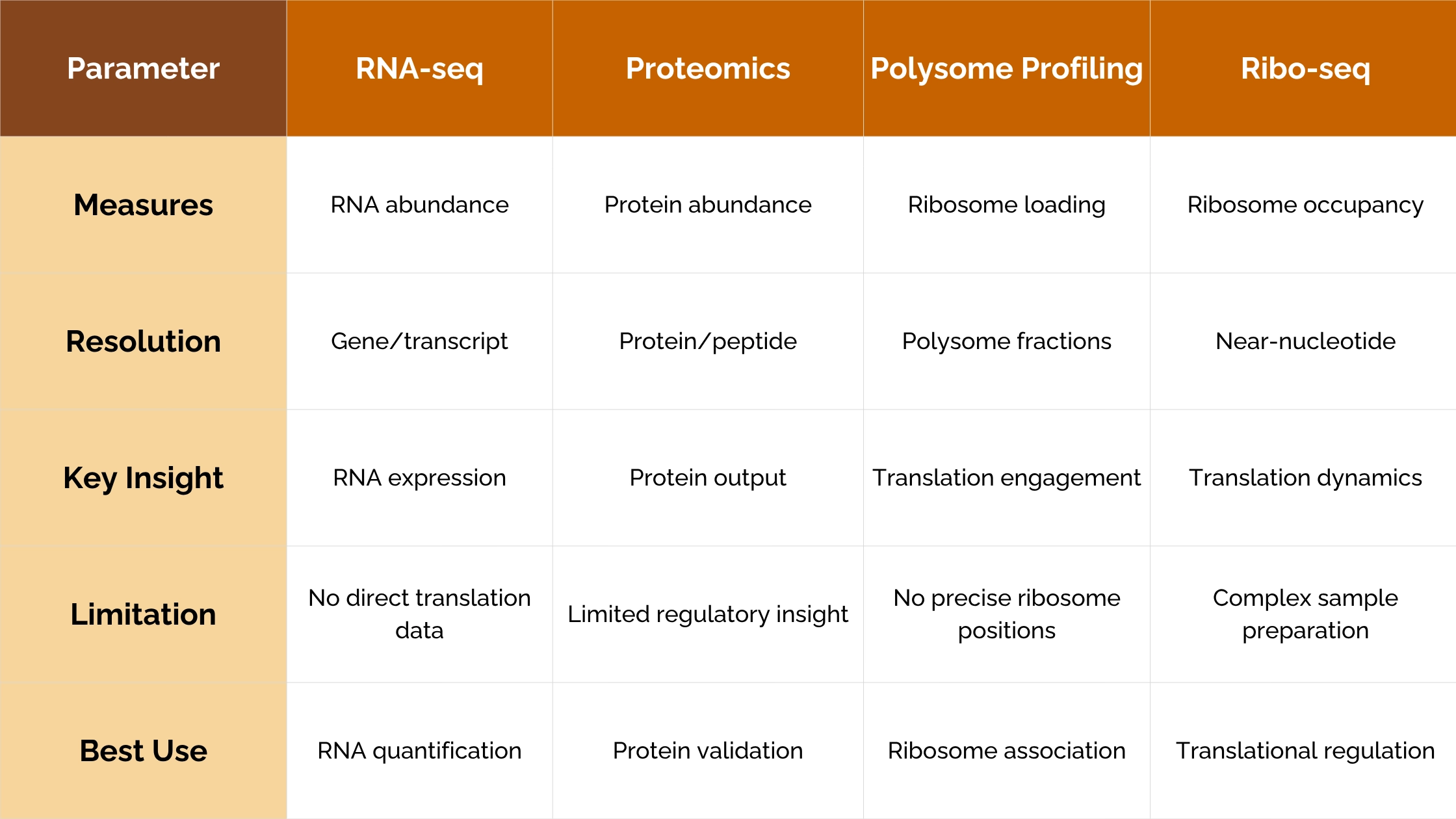
RNA-seq measures
transcript abundance but does not directly determine whether a transcript is
being translated. Protein assays and proteomics measure downstream output but
may not identify where translational regulation occurs. Polysome profiling can
show the association of transcripts with multiple ribosomes, while ribosome
profiling provides much finer positional information across translated RNAs
[2].
When
RNA-seq and Ribo-seq are analyzed together, researchers can compare transcript
abundance with ribosome-protected fragment abundance to estimate relative
translation efficiency.
Comparing translation efficiency between conditions
Calculating TE for one sample is straightforward; deciding whether TE has genuinely changed between conditions is harder. The deltaTE (ΔTE) approach developed by Chothani, Rackham and colleagues models matched RNA-seq and Ribo-seq counts together, so that complex experimental designs and confounders such as batch can be accounted for, and it outperformed the other methods tested in their benchmarking
ΔTE also classifies genes by regulatory mode: exclusive genes change in ribosome occupancy and TE without a change in mRNA; forwarded genes change in mRNA and ribosome occupancy together, with no change in TE; buffered genes show a TE change that counteracts the change in mRNA; and intensified genes show a TE change that amplifies it. For a therapeutic program, this distinction helps separate effects on transcription or RNA stability from effects on translation itself.
Data quality matters as much as the statistics. Ribo-seq data are sparse, coverage along open reading frames is not uniform, and codon-level factors such as tRNA availability can locally raise or lower footprint density.
For RNA therapeutics, this can be particularly informative when two constructs show similar RNA levels but different protein output. Researchers can then investigate whether the difference appears at ribosome engagement, initiation, elongation or another translational step.
How ribosome profiling supports RNA therapeutic development
Ribosome profiling for RNA therapeutics provides a view of ribosome position across translated transcripts.
It can help researchers investigate whether one candidate is more strongly translated than another, whether ribosomes accumulate at particular regions, whether a coding-sequence change alters translation behavior, or whether a treatment causes translational changes across the wider transcriptome [2]. CSH Perspectives in Biology
Ribo-seq can therefore support candidate comparison, mechanism-of-action studies and the investigation of differences between RNA abundance and protein output.
From ribosome profiling to active translation
Conventional Ribo-seq profiles ribosome-protected fragments across transcripts. RiboLace was developed as a complementary approach that enriches actively translating ribosomes through an antibody-free and tag-free pull-down using a puromycin-derived molecule [7].
The original RiboLace study reported positional information on active ribosomes at nucleotide resolution, translation-level estimates and applicability to low amounts of input material [7]. PubMed
This makes active ribosome enrichment particularly relevant when the research question requires a closer examination of the active translatome.
What low input translatomics can reveal
A useful example comes from human oocyte maturation, where sample availability makes conventional approaches difficult.
Hu and colleagues developed T&T-seq, combining transcriptome sequencing with RiboLace-based translatome profiling in low-input and single-oocyte samples [8].
In their experimental comparison, T&T-seq translatome mapping rates averaged 91.45% for 10-oocyte samples and 79.29% for single-oocyte samples, while their miniRibo-seq workflow reached 21.60% [8]. These figures describe that specific experimental comparison and should not be interpreted as universal performance values for Ribo-seq. Nature
The biological result was equally interesting.
Principal-component analysis of human transcriptomes showed that immature GV and mature MII oocytes partially overlapped. In contrast, the translatome profiles separated GV and MII oocytes more clearly, indicating a more distinct expression pattern at the translational level [8]. Nature
The study also identified OOSP2 as an oocyte maturation factor and investigated transcriptional and translational responses following OOSP2 treatment. For specific pathways, including small GTPase signaling and organelle localization, transcriptional levels remained similar between control and OOSP2-treated oocytes while translational levels increased strongly [8]. Nature
This illustrates a key value of translatomics: biologically meaningful regulation can sometimes become clearer at the level of translation than at the level of transcript abundance.
Using translational data to optimize therapeutic mRNA
A practical mRNA optimization strategy should begin with the biological question, not a single design rule.
Candidate RNAs can first be evaluated for abundance and stability. Translation-level measurements can then show how those transcripts interact with ribosomes. If differences appear, researchers can investigate factors such as UTR architecture, initiation, coding sequence, RNA structure, codon choice or ribosome pausing. Protein measurements can then confirm the final functional outcome.
A useful development workflow is:
Design → Measure RNA → Measure Translation → Identify Bottlenecks → Redesign → Validate Protein Output
This approach creates a clearer link between sequence design and functional protein production.
Beyond RNA abundance
RNA therapeutics increasingly rely on sophisticated sequence design, delivery systems and chemical modifications. But once an mRNA reaches the cell, its functional performance still depends on how effectively the translation machinery reads it.
mRNA translation efficiency should therefore be considered more than a downstream validation metric. It can provide an additional decision-making layer for candidate comparison, sequence optimization and mechanism-of-action studies.
RNA-seq tells us whether a transcript is present.
Ribosome profiling helps reveal whether, where and how that transcript is being translated.
For therapeutic mRNA development, understanding that difference can provide information that transcript abundance alone cannot.
Immagina Biotechnology supports ribosome profiling and active translatome analysis for researchers who need to investigate translation beyond RNA abundance.
Want to understand how efficiently your mRNA candidates are being translated? Explore how ribosome profiling and active translation analysis can support your RNA therapeutic development strategy.
References
[1] Yang B, Sedano MJ, Perez K, et al. RNA therapeutics: current status and future directions. Signal Transduction and Targeted Therapy. 2026;11:409. DOI: 10.1038/s41392-026-03032-5. Nature
[2] Ingolia NT, Hussmann JA, Weissman JS. Ribosome Profiling: Global Views of Translation. Cold Spring Harbor Perspectives in Biology. 2019;11(5):a032698. DOI: 10.1101/cshperspect.a032698. PubMed
[3] Zheng D, Persyn L, Wang J, et al. Predicting the translation efficiency of messenger RNA in mammalian cells. Nature Biotechnology. 2026;44:783–796. DOI: 10.1038/s41587-025-02712-x. Nature
[4] Lewis CJT, Xie LH, Bhandarkar SM, et al. Quantitative profiling of human translation initiation reveals elements that potently regulate endogenous and therapeutically modified mRNAs. Molecular Cell. 2025;85(2):445–459.e5. DOI: 10.1016/j.molcel.2024.11.030. PubMed
[5] Li Y, Wang F, Yang J, et al. Deep generative optimization of mRNA codon sequences for enhanced mRNA translation and therapeutic efficacy. Nature Communications. 2025;16:9957. DOI: 10.1038/s41467-025-64894-x. DOI.org
[6] Leppek K, Byeon GW, Kladwang W, et al. Combinatorial optimization of mRNA structure, stability, and translation for RNA-based therapeutics. Nature Communications. 2022;13:1536. DOI: 10.1038/s41467-022-28776-w. Nature
[7] Clamer M, Tebaldi T, Lauria F, et al. Active Ribosome Profiling with RiboLace. Cell Reports. 2018;25(4):1097–1108.e5. DOI: 10.1016/j.celrep.2018.09.084. PubMed
[8] Hu W, Zeng H, Shi Y, et al. Single-cell transcriptome and translatome dual-omics reveals potential mechanisms of human oocyte maturation. Nature Communications. 2022;13:5114. DOI: 10.1038/s41467-022-32791-2. Nature